Select your language
Select your language
")
")
Web site:
Products:
Why Ridet?
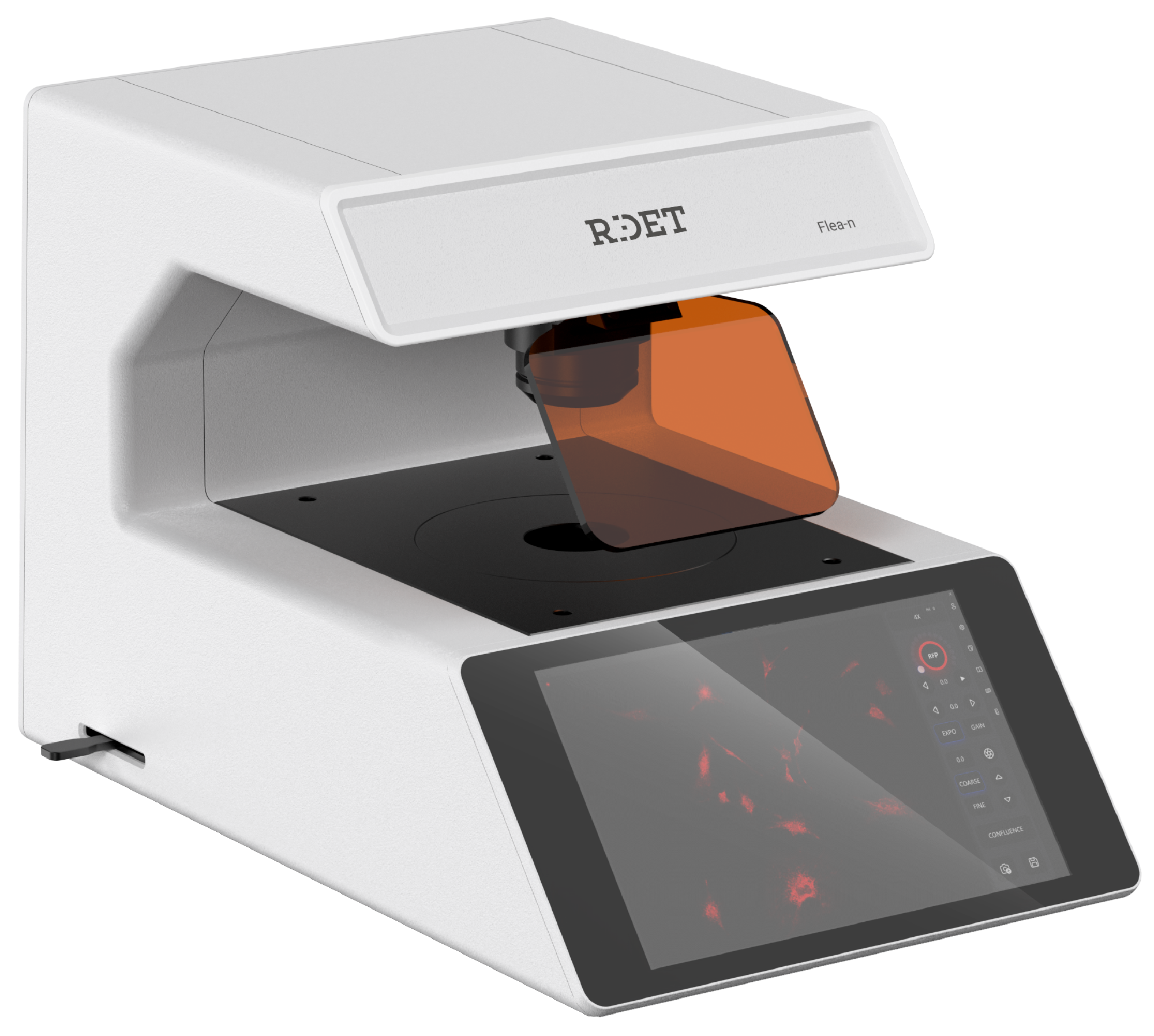
Flea-N
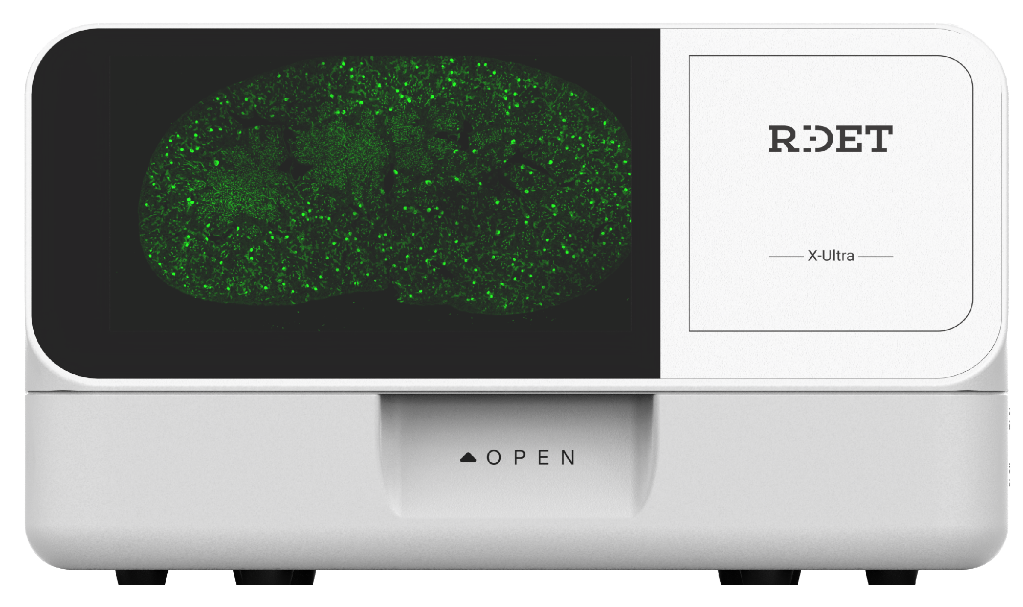
X-Ultra
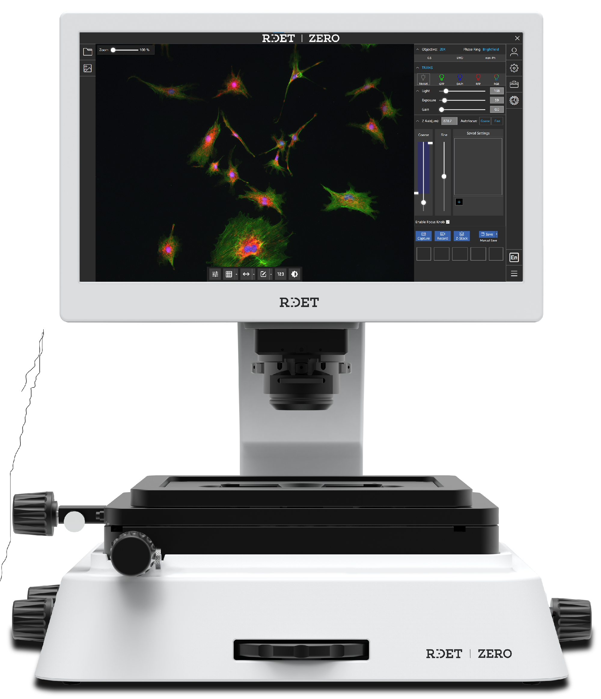
ZERO
Click to enlarge
We use cookies and similar technologies for site functionality (essential) and analytics (non-essential). By clicking "Confirm", you consent to our use of these technologies as described in our Privacy Policy, which includes details on how to manage or withdraw consent.
Pro zajištění funkčnosti webu a pro analýzu provozu používáme soubory cookie a podobné technologie. Kliknutím na „Confirm“ souhlasíte s jejich používáním tak, jak je popsáno v našich Zásadách ochrany osobních údajů, které zahrnují i informace o odvolání souhlasu.